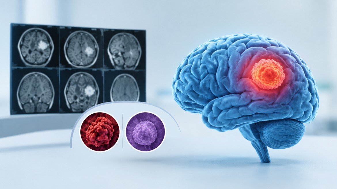
Ask ten people what a brain tumor is and you’ll get one picture: a lump, a scary one, probably fatal. The reality is messier. There are over a hundred recognised tumor types, and some of them will never need an operation.
Glioblastoma is the one people tend to have in mind when they hear the phrase, and its reputation is earned. But if you’re holding a pathology report, or searching for a neurosurgeon in Noida or a brain tumor surgeon in Noida to explain one, it helps to know where glioblastoma sits among its neighbours.
Boulders and Weeds
Here is the analogy I find most useful.
Some tumors behave like a boulder in a garden bed. They press on the soil around them but keep a clear edge. Dig carefully and you can often lift the whole thing out.
Others behave like weeds. Their roots run through the soil, so pulling everything visible still leaves fine roots behind.
Glioblastoma is the weed. Its cells migrate into healthy brain tissue beyond what any scan shows. That single trait explains why surgery is only the first step of its treatment.
Glioblastoma in Plain Terms
It starts in glial cells, which support and feed neurons. The World Health Organization classes it as grade 4, the highest. Current classification names it glioblastoma, IDH-wildtype, meaning it lacks a gene mutation seen in some slower gliomas.
It is the most common malignant primary brain tumor in adults, and it grows fast. Symptoms often build over weeks: worsening headaches, a first seizure, speech trouble, weakness on one side, or a change in personality that family members notice before the patient does.
Standard care combines surgery, radiation, and temozolomide chemotherapy. In the landmark trial that established this approach, median survival was about 15 months. Some people live much longer. That figure describes a trial population, not any individual, and age, general health, how much tumor is removed, and tumor genetics all shift the picture.
The Rest of the Family
Meningioma. Grows from the membranes around the brain, not the brain itself. Most are grade 1 and slow. Many are simply watched. When surgery is needed, full removal is often realistic.
Low-grade glioma. Same cell family as glioblastoma, far slower pace. Common in younger adults, often discovered after a seizure. They infiltrate, so cure is harder than with a meningioma, but years of stability are common. Some progress to higher grades later.
Pituitary adenoma. Mostly benign, found at the base of the brain.These tumors may cause hormonal imbalances or put pressure on the optic nerves, leading to vision problems. Many of these tumors can be removed through the nasal passage, avoiding the need for an incision in the skull.
Vestibular schwannoma. A benign growth on the hearing and balance nerve. Options are monitoring, radiosurgery, or surgery.
Metastases. Worth knowing: in adults, tumors that have spread to the brain from lung, breast, or other cancers are more common than primary brain tumors. Their treatment follows the original cancer.
Side by Side
| Feature | Glioblastoma | Meningioma | Low-grade glioma | Pituitary adenoma |
| Starts in | Glial cells | Brain membranes | Glial cells | Pituitary gland |
| WHO grade | 4 | Mostly 1 | 1–2, some higher | Generally benign |
| Speed | Fast | Usually slow | Slow | Usually slow |
| Edge | Poorly defined | Usually clear | Often blurred | Usually clear |
| Complete removal | Rarely | Often | Sometimes | Often |
| After surgery | Radiation plus chemotherapy | Observation, sometimes radiation | Monitoring or further therapy | Medication, monitoring, or both |
The Scan Suggests, the Pathology Decides
An MRI can point toward a diagnosis. Only tissue examined under a microscope, along with molecular testing, confirms it. Three markers appear often in reports:
- IDH mutation: usually goes with slower gliomas and a better outlook
- MGMT methylation: suggests better response to temozolomide
- 1p/19q co-deletion: identifies oligodendroglioma, which tends to respond well to treatment
Two tumors that look alike on film can carry very different genetics. That is why a tissue sample matters even when the scan seems obvious.
What the Process Usually Looks Like
- MRI with contrast to map size and position.
- A surgical decision based on how much can be removed without costing function.
- Pathology and molecular testing on the tissue.
- Radiation, chemotherapy, or both if the diagnosis calls for it.
- Scheduled follow-up scans for regrowth.
A meningioma may skip steps four and five. For glioblastoma they carry most of the plan.
Questions Worth Bringing to the Consultation
Brain tumor care involves neurosurgery, radiology, pathology, and oncology working together. When you meet the surgeon, consider asking:
- What type of tumor do you suspect, and what will confirm it?
- How much can be removed safely, and what function is at risk?
- Will my case be discussed with oncology and radiation colleagues?
In India, a practising doctor should be registered with a State Medical Council, and the National Medical Commission’s National Medical Register lets you verify registration and qualifications. Be cautious of anyone who guarantees results. Honest uncertainty is usually a sign of experience.
Frequently Asked Questions
Is glioblastoma the same as brain cancer?
It is one type, and the most common malignant primary one in adults. “Brain tumor” also covers benign growths and spread from other cancers.
Can it be removed completely?
The visible mass can be removed, but microscopic cells typically remain, which is why radiation and chemotherapy follow.
Are all meningiomas harmless?
Most are benign. A minority are higher grade, and even benign ones can cause problems depending on size and location.
Does every brain tumor need surgery?
No. Small, slow, symptom-free tumors are often monitored.
The Final Word
Glioblastoma differs from other brain tumors because it can grow rapidly, spread into nearby healthy brain tissue, and respond differently to treatment. A quiet meningioma and a grade 4 glioma share a label and little else. Whatever the diagnosis, the exact type, confirmed by pathology and molecular testing, drives every later decision. If you are choosing a neurosurgeon in Noida or a brain tumor surgeon in Noida, look for someone who explains your diagnosis plainly, brings in a wider team, and is honest about what can’t be promised.
Dr. Rahul Gupta is Senior Director and Head of Neurosurgery at Fortis Hospital, Noida, with over 20 years of experience in the assessment and management of brain, spine, and nerve disorders. His clinical practice includes minimally invasive brain and spine surgery, brain tumour management, neurovascular procedures, and other neurosurgical treatments. Treatment recommendations are based on each patient’s diagnosis, medical history, and individual clinical needs.