You know the moment. You open the MRI report app, or someone hands you a folder, and there it is — a paragraph full of words like “desiccation,” “disc bulge,” “mild canal stenosis,” and your first instinct is to assume the worst. I’ve watched this happen to more people than I can count: they read a radiology report before ever discussing it with a doctor, and by the time they walk into a consultation, they’ve already convinced themselves surgery is coming. Most of the time, it isn’t. Understanding what these terms actually mean is the difference between reasonable concern and unnecessary panic, and it’s usually the first real step before anyone talks about spine treatment in Noida.
What Is Cervical Disc Disease?
The cervical spine contains seven vertebrae. Between most of these bones are soft intervertebral discs that act as cushions and allow the neck to move.
With age, discs can gradually lose water and become less flexible. Small tears, disc bulging, loss of disc height and changes in nearby joints may develop. These are commonly described as degenerative disc disease or cervical spondylosis.
These changes can occur without causing symptoms. In fact, degenerative cervical disc findings are common even in people who don’t have neck pain. One review cited by the American Academy of Family Physicians found degenerative cervical discs on MRI in more than 85% of asymptomatic people over 65.
That is why an MRI report needs to be interpreted alongside the person, not separately from the person.
Let’s Decode a Real Report, Line by Line
Picture a fairly typical cervical MRI report. It might read something like: “Disc desiccation at C5-C6 with mild posterior disc bulge. Mild flattening of the thecal sac. No significant central canal or foraminal stenosis.”
Here’s what that actually translates to in plain language.
Disc desiccation just means the disc has lost some water content — which happens to basically everyone as they age, starting sometimes as early as the late twenties. It’s not a diagnosis of anything painful on its own. Think of it as the spinal equivalent of gray hair. Nearly universal, rarely the actual problem.
Disc bulge describes the disc’s outer layer extending slightly beyond its normal boundary, in every direction, like a tire that’s lost a bit of its shape without actually tearing. A bulge is different from a herniation — a herniation means part of the disc’s inner material has pushed through a tear in the outer layer, which is a more specific, sometimes more significant finding.
Thecal sac flattening sounds alarming but often isn’t. The thecal sac is the membrane surrounding the spinal cord and fluid around it. Mild flattening means something — a bulging disc, usually — is touching that sac slightly. It doesn’t necessarily mean the spinal cord itself is compressed or damaged.
No significant stenosis is actually the reassuring part of that report, and it’s easy to skip past it while fixated on the scarier-sounding terms above it. Stenosis means narrowing of the space around the spinal cord or nerve roots. If there’s none, or only mild narrowing, that’s genuinely good news.
Why Your MRI Might Look Worse Than It Feels
This is the part that confuses almost everyone. A landmark study published years ago scanned the necks of people with zero neck pain and found that a large share of them — well over half in some age groups — had disc bulges or degeneration visible on MRI despite feeling completely fine.
What this tells us: the spine changes with age whether or not it hurts. An MRI is a snapshot of anatomy, not a measurement of pain. This is exactly why a good specialist won’t treat a scan — they treat the actual symptoms a person is experiencing, cross-checked against what the imaging shows.
When the Findings Actually Matter
Not every abnormal finding is irrelevant, though. A few things genuinely shift the picture.
- Nerve root compression correlating with symptoms. If the MRI shows a disc pressing on a specific nerve root, and that lines up with pain, numbness, or weakness radiating down the same arm, that’s a meaningful match worth acting on.
- Spinal cord compression (myelopathy). This is more serious. Signs include changes in hand coordination, difficulty with fine motor tasks like buttoning a shirt, or balance problems while walking. Cord compression visible on MRI alongside these symptoms usually moves the conversation toward more active treatment.
- Progressive weakness. A muscle that’s getting weaker over weeks, not just painful, points toward nerve involvement that needs a closer look rather than a wait-and-see approach.
What Typically Happens After Diagnosis
Step One: Match the Scan to the Symptoms
A specialist worth trusting spends real time asking questions before jumping to the imaging — where exactly the pain radiates, whether it follows a specific pattern down the arm, what makes it worse or better, how long it’s been going on. The MRI supports this conversation; it doesn’t replace it.
Step Two: Conservative Treatment, Almost Always First
Physical therapy, targeted neck exercises, short courses of anti-inflammatory medication, and sometimes a cervical collar for brief periods during acute flare-ups. Most cervical disc issues improve substantially within six to twelve weeks with this approach alone.
Step Three: Escalating Only When Needed
If pain persists well beyond a reasonable trial period, or there’s measurable weakness, options like targeted epidural injections come next. Surgery — usually a discectomy, sometimes with fusion — becomes relevant mainly when conservative treatment hasn’t worked, or when there’s clear nerve or spinal cord compression causing ongoing damage.
What Does Cervical Disc Disease on MRI Actually Show?
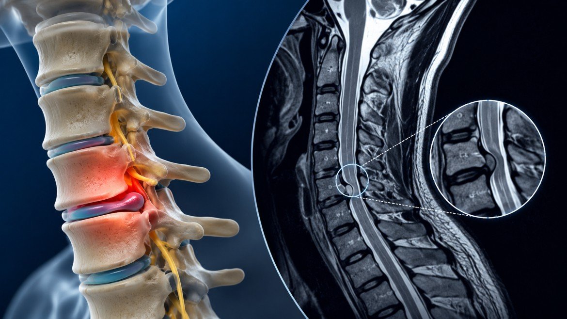
An MRI gives doctors a detailed view of the discs, spinal cord, nerve roots and surrounding soft tissues.
Here are some of the terms you may see in a report:
| MRI Term | What It Generally Means |
| Disc degeneration | The disc has developed age- or wear-related structural changes |
| Disc bulge | The disc extends beyond its usual boundary over part of its circumference |
| Disc protrusion | A more focal disc displacement |
| Disc herniation | Disc material extends beyond the normal disc space and may affect nearby structures |
| Foraminal narrowing | The opening through which a nerve exits has become narrower |
| Spinal canal stenosis | The space available for the spinal cord or nerves has narrowed |
| Cord compression | The spinal cord is being pressed or deformed |
| Osteophytes | Bone spurs that can develop with degenerative changes |
These findings don’t all carry the same clinical significance.
For example, a small disc bulge without nerve compression may have little connection with someone’s symptoms. A disc herniation that compresses a nerve root and matches the patient’s arm pain, numbness or weakness is more clinically relevant.
Finding the Right Spine Treatment in Noida
If you’re searching for Spine Treatment in Noida, don’t choose a treatment solely because an MRI report contains words such as “severe,” “degenerative,” or “protrusion.”
A useful consultation should connect three things:
Symptoms → examination → imaging
That connection helps determine whether the MRI finding is actually responsible for the problem and whether treatment should be conservative, interventional or surgical.
A second opinion can also be reasonable when surgery has been suggested, particularly if the MRI findings and symptoms don’t seem to match.
Frequently Asked Questions
Does a disc bulge always mean I’ll eventually need surgery?
No. Most disc bulges never require surgery and improve with conservative treatment. Surgery becomes relevant mainly when there’s significant nerve compression causing persistent symptoms that haven’t responded to other treatment.
Why does my neck hurt if the MRI shows only mild findings?
Pain and imaging findings don’t always correlate closely. Muscle strain, posture-related tension, and other soft tissue issues can cause real pain even when the disc findings themselves look fairly minor.
How often should I get a follow-up MRI?
Unless symptoms are worsening or new neurological signs appear, repeat imaging usually isn’t necessary. Most specialists reassess based on how symptoms are actually changing, not on a fixed imaging schedule.
Is cervical disc disease the same as a slipped disc?
Not quite. “Slipped disc” is a general term people use loosely, but the disc doesn’t actually slip out of place — it either bulges or herniates, which are more precise descriptions of what’s happening structurally.
Reading your own MRI report doesn’t have to mean spiraling into worst-case assumptions. Most findings described in these reports are simply part of normal spinal aging, and only a smaller subset actually needs active treatment. If your symptoms and imaging genuinely line up toward something requiring intervention, proper spine treatment in Noida means working with a specialist who reads the whole picture, not just the scarier-sounding lines in a radiology report.
Conclusion
Cervical disc disease shown on MRI does not automatically mean a problem or that surgery is needed. Disc degeneration, bulging and narrowing are findings especially as people age. What matters is whether abnormality matches your symptoms and whether abnormality is seen in your exam.
If you’re considering Spine Treatment in Noida, the most useful next step is not to treat the MRI report alone. Have the images and report reviewed alongside your symptoms and examination so that the treatment plan addresses the actual cause of your problem.

Dr. Rahul Gupta is Senior Director and Head of Neurosurgery at Fortis Hospital, Noida, with over 20 years of experience in the assessment and management of brain, spine, and nerve disorders. His clinical practice includes minimally invasive brain and spine surgery, brain tumour management, neurovascular procedures, and other neurosurgical treatments. Treatment recommendations are based on each patient’s diagnosis, medical history, and individual clinical needs.